
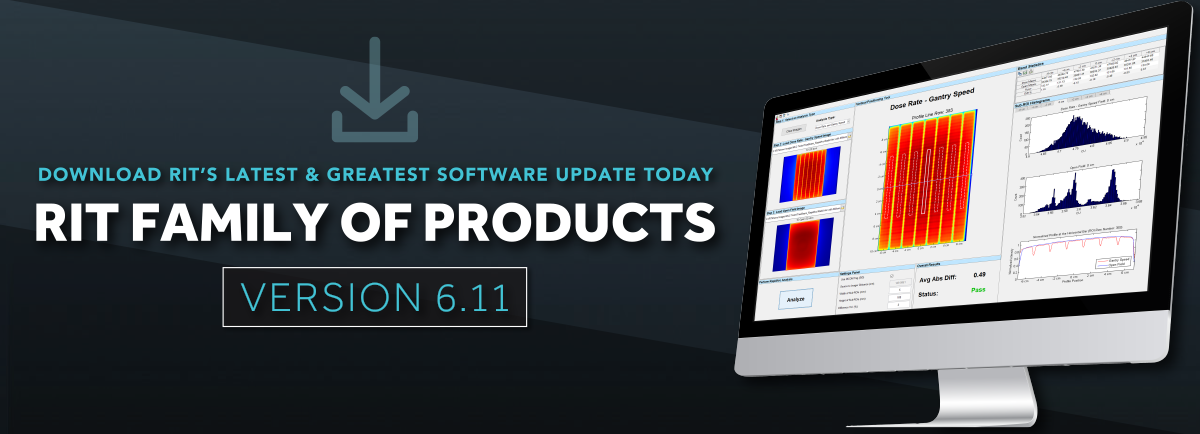

An impressive range of products
Radiological Imaging Technology
Machine QA MLC QA Patient QA Imaging QA
Imaging QA/QC
RIT offers a range software packages for automated imaging phantom analysis, from specific routines following task group recommendations, to a full suite of one-click, instant analyses of therapeutic and diagnostic images. All products come equipped with tracking, trending and automation tools designed to optimize your workflow.
Radia Modules
Radiological Imaging Technology® offers Radia software: an à la carte-based software for comprehensive imaging QA/QC at your facility. Easily customize your Radia software by adding the diagnostic and/or therapy modules needed to perform automated analyses on your facility's phantoms. Radia is tailored specifically to you - you can select and purchase any of the modules listed below to create a customized software package for your imaging QA/QC needs:
- STEP 1:
If you do not have an existing RIT product with Imaging QA/QC (RIT Complete, RITG142, or RITG148+) you will be required to purchase the Radia base module, which offers basic imaging QA/QC functionality and includes our automated features like RITtrend, RunQueueC, and Cerberus.
- STEP 2:
Select the Radia modules you need à la carte, based on the automated phantom analyses needed at your facility. Radia software packages can be fully customized with any of the 14 modules listed below for therapy and/or diagnostic imaging QA/QC.
| Features | Details |
|---|---|
| Locate & Import Images | DICOM directory browser, open directory browser, DICOM file filter for organizing your images |
| Output Formats | Print, PDF, Excel (with custom templates) |
| DICOM Image Viewer & DICOM Tag Viewer | View DICOM images and DICOM tags |
| Manual Measurements | Distance, angle, profile, histograms, round and square ROI. ASCII output of profiles. |
| Monitor Image Quality | SMPTE pattern and TG18 images with custom templates (print, PDF, Excel) |
 |
Batch processing of images, including preference and tolerance profile customization. |
 |
Monitors folders and processes incoming files without you lifting a finger or even being present. Up to 4 DICOM tags may be used to sort. |
 |
All measurements can be tracked and plotted over time in the statistical trend database. Any number of databases can be used. All Statistical Process Control parameters measured and plotted. |
| QA/QC Type | Module Name | Phantoms for Analysis | Analysis Measurements |
|---|---|---|---|
| Diagnostic | CT Uniformity Module | Generic CT Water Phantoms | Uniformity, mean pixel value and SD by measurement position |
| GE CT Module | Optima 660 Phantom | Contrast, CNR, slice thickness, alignment accuracy, MTF, modulation, low contrast, noise, and uniformity | |
| IEC 62464 MRI Module | IEC 62464 MRI Phantom | Uniformity, ghosting, contrast, CNR, geometric distortion, modulation, MTF, and slice thickness | |
| ACR Small MRI Module | ACR Small MRI Phantom (Extremity & Breast) | Localizer line length, geometric accuracy, resolution, slice thickness, slice difference, geometric distortion, uniformity, ghosting, low contrast: spokes detected, and contrast to noise ratios | |
| ACR FFDM Module | Gammex FFDM Phantom | Insert width, contrast analysis, and insert analysis (fibers, specs, and masses) | |
| CIRS FFDM Phantom | Insert width, contrast analysis, and insert analysis (fibers, specs, and masses) | ||
| Gamma Camera Uniformity Module | None (Flood field) | NEMA NU 1-2012: Useful field of view uniformity measurements and statistics, useful field of view defective pixels, central field of view uniformity measurements and statistics, and central field of view defective pixels |
| QA/QC Type | Module Name | Phantoms for Analysis | Analysis Measurements |
|---|---|---|---|
| Diagnostic & Therapy | ACR CT Module | ACR CT Phantom | Landmark BB position, Landmark slice thickness, CT material values, CT material linearity, uniformity, resolution, and MTF |
| ACR MRI Module | ACR Large MR Phantom | Localizer line length, geometric accuracy, resolution, slice thickness, slice difference, geometric distortion, uniformity, ghosting, low contrast: spokes detected, and contrast to noise ratios | |
| kV Imaging Module | IBA Primus®-L Phantom | Noise and CNR for each step in the step wedge, CNR by contrast object, ytterbium indicator, central area uniformity, resolution line pairs detected, MTF, and geometry. Couch placement analysis is supported. | |
| Leeds TOR 18FG Phantom | Contrast to noise ratio for high contrast and low contrast objects, resolution line pairs detected, modulation. Couch placement analysis is supported. | ||
| PTW NORMI® 4 Phantom (20 x 20 cm and 30 x 30 cm) | Noise and CNR for each step in the step wedge, CNR by contrast object, ytterbium indicator, central area uniformity, resolution line pairs detected, MTF, and geometry. Couch placement analysis is supported. | ||
| Standard Imaging QCkV-1 Phantom | Contrast value, noise value, uniformity (NEMA), spatial resolution, modulation. Couch placement analysis is supported. | ||
| Catphan/OBI Module | CATPHAN® 500/600 Series Phantom | CT material values, CT number linearity, slice width (both horizontal and both vertical slices), ramp alignment, ramp yaw and tilt, rod distances (geometric distortion), low contrast spheres, contrast to noise ratio, CT uniformity, integral uniformity, resolution, MTF | |
| Elekta 503 CATPHAN® (XVI) Phantom | Spatial resolution, MTF (based on standard deviation), MTF curve, uniformity, sagittal geometry, low contrast, horizontal and vertical scaling, material values for LDPE and Poly | ||
| Varian 504 & 604 CATPHAN® Phantom | CT material values, CT number linearity, slice width (both horizontal and both vertical slices), ramp alignment, ramp yaw and tilt, rod distances (geometric distortion), low contrast spheres, contrast to noise ratio, CT uniformity, integral uniformity, resolution, MTF | ||
| Siemens MV CT (OBI) Phantom | Geometric analysis (BBs), high and low contrast objects, resolution line pairs detected, MTF (based on standard deviation), and uniformity |
Catphan® is a registered trademark of The Phantom Laboratory.
NORMI® is a registered trademark of PTW.
Primus® is a registered trademark of IBA.
| QA/QC Type | Module Name | Phantoms for Analysis | Analysis Measurements |
|---|---|---|---|
| Therapy | Electron Density/Tissue Characterization Module | Accuray TomoTherapy® Cheese MVCT Phantom | Materials measurements, CNR, resolution, noise, and uniformity |
| CIRS CT Phantoms: 062, 062A, 062M (Cone Beam) | CT material value measurements | ||
| Gammex 467 CT Phantom | CT material value measurements, rod distance measurements | ||
| EPID MV Imaging Module | RIT EPID Phantom (EPID QA Phantom - manufactured and sold by Standard Imaging.) | Geometric accuracy, uniformity area, contrast (AI plugs), copper plug isotrophy, resolution (MTF). Offers support for all EPID devices: Varian aS500 / aS1000, Elekta iView™ and Siemens. Couch placement analysis is supported. | |
| PTW EPID QC Phantom | Signal linearity, SNR, response linearity, isotropy, geometric accuracy, MTF, low contrast measurements. Offers support for all EPID devices: Varian aS500/aS1000, Elekta iView™ and Siemens. Couch placement analysis is supported. | ||
| Las Vegas EPID Phantoms | Contrast to noise ratios, spatial resolution, Varian image quality test, and background statistics, TG-58 contrast and spatial resolution test. Includes added TG-58 and Varian QA acceptance criteria for analysis. Couch placement analysis is not supported. | ||
| Standard Imaging QC-3 Phantom | Contrast value, noise value, uniformity (NEMA), line pairs detected, modulation. Couch placement analysis is supported. | ||
| Penta-Guide Module | Modus (IBA) Penta-Guide Phantom | DRR-MV-kV coincidence, CBCT axial and sagittal positions | |
| ISOCube Module | Standard Imaging ISOCube Phantom | kV-MV coincidence, CBCT isocenter alignment, kV Collimation, MV – Radiation/Light Field coincidence, and 6 degree-of-freedom couch measurements |
TomoTherapy® is a registered trademark of Accuray, Inc.
Product Packages
The RIT product packages in the table below feature Imaging QA/QC capabilities. The modules in Radia (both Therapy and Diagnostic) require a Radia Base Module, but all other modules can be fully customized à la carte. You can mix and match Radia's therapy and diagnostic modules. The other product packages, RIT Complete, RITG142, and RITG148+, do not require a Radia Base module, as they have built-in imaging QA capabilities. The modules featured in RIT Complete, RITG142, and RITG148+ are included within the package as listed, but additional therapy and diagnostic modules can be added to existing packages.
| Module Name | Radia (Diagnostic) Modules are optional, can be customized, and added à la carte. | Radia (Therapy) Modules are optional, can be customized, and added à la carte. | RIT Complete Modules are included within package as listed. | RITG142 Modules are included within package as listed. | RITG148+ Modules are included within package as listed. |
|---|---|---|---|---|---|
| ACR CT | ✔ | ✔ | |||
| ACR FFDM | ✔ | ||||
| ACR MRI | ✔ | ✔ | |||
| ACR Small MRI |
✔ | ||||
| Catphan/OBI | ✔ | ✔ | ✔ | ✔ | |
| CT Uniformity |
✔ | ||||
|
Electron Density/Tissue Characterization |
✔ | ✔ | ✔ | ||
| EPID MV Imaging |
✔ | ✔ | ✔ | ||
| GE CT (Optima 660) |
✔ | ||||
| Gamma Camera Uniformity |
✔ | ||||
| IEC 62464 MRI |
✔ | ||||
| ISOCube | ✔ | ✔ | ✔ | ||
| kV Imaging |
✔ | ✔ | ✔ | ✔ | |
| Penta-Guide | ✔ | ✔ | ✔ | ||
| Radia Base |
Required | Not Required |
|||
Automated Features
Cerberus: Hands-Free, Automated Phantom Analysis
The RIT Cerberus system constantly operates in the background of your machine, without even having to open RIT software. Automatically monitor folders and pinpoint specific files to process and analyze, matching any set criteria, such as file naming patterns, DICOM tag matches, or file types. Cerberus allows you to sort with up to four DICOM tags. It will then use these DICOM tags to select and process incoming files. Cerberus will automatically perform analyses, generate reports, and share data to RITtrend™ for statistical database analysis.

RunQueueC: One-Click Automated Batch Analysis
Fast, consistent batch phantom analysis with a single click
RunQueueC is a feature included in all software packages with imaging QC capabilities. This includes RIT Complete, RITG142, RITG148+, Radia Therapy, and Radia Diagnostic. RunQueueC performs one-click batch analysis on any number of images are queued into the system. This feature is very useful for:
- Simultaneously running all modules of a CBCT scan (e.g. CATPHAN®), as pictured in the workflow illustration below.
- Processing MV/kV images with a single click.
- Saving time, with no need to sift through a stack of DICOM images to look for a specific image to analyze.
- Analyzing troves of archived data and instantaneously adding them to RITtrend™ for tracking and trending.

1. One-Time Setup: Create reusable analysis protocols, checklists, and reporting scripts for your imaging QC.
2. Execute RunQueueC: Select a large set of images to create a batch. Next, execute one of the pre-established protocols to automatically-generate reports.
3. Automatic Analysis: In seconds, each image slice of the phantom will be analyzed automatically and the results exported to your preferred format.
The graphic above illustrates the simplified workflow when using RunQueueC to perform batch analysis on images from the CATPHAN® phantom. After you acquire entire stack of CATPHAN® DICOM images, you will specify the folder in RunQueueC and the images will be batch-processed for analysis at once. There is no need to manually pick out each, individual image and process separately, completely streamlining your imaging QC workflow.

Finally, after the analysis is completed, you can select the format for the exporting the results. Reports can be generated in the form of PDFs, Excel spreadsheets, or uploaded to the RITtrend™ SPC database for statistical tracking and trending. Preference profile customization has been added to RunQueueC to increase convenience and efficiency for our users. Your preference and tolerance profiles can be loaded directly from the interface, without having to open the Profile Manager. This allows you to easily select a profile for a customized analysis report specific to your machine.
CATPHAN® is a registered trademark of The Phantom Laboratory.
Tolerance Management

- Conveniently set and toggle between tolerances in an easy-to-use panel, located in the main interface, and displayed in every phantom analysis routine.
- Set your own PASS/FAIL criteria for analyses and their corresponding reports, allowing for complete customization in all imaging QA analyses.
- Preference profiles and tolerances are set on a
per-machine basis. They can be precisely-tailored to each individual machine in use.
Tolerance Profile Management
Set preference profiles for your tolerance measurements on a per-machine basis. Preference profiles can be precisely-tailored to each individual machine in use. In the illustration, we see that there are two users: User A and User B, managing three different machines, each with a specific preference profile: LINAC #1, LINAC #2, and CT#1. User A can manage LINAC #1 and User B can manage CT#1, but both User A & User B can manage LINAC #2. Tolerance preference profiles offer versatility when managing multiple machines at a facility.
Conveniently toggle between tolerance preference profiles using the pull-down menu in the main window of the RIT software interface.

Integration with RITtrend ™
Transfer all PASS/FAIL tolerance data to the RITtrend™database for statistical tracking and trending.
- Easily import your analysis results into the database with a single click.
- Set baseline measurements for statistical process control.
- Track historical data over time to detect any anomalies in your measurements.
- Enable alerts via email, allowing you to immediately view significant changes in data or measurements that may exceed their set tolerances.
RITtrend™is a registered trademark of Radiological Imaging Technology, Inc.

Analysis Results

Imaging QA analysis results go into deeper detail, specifying both the status of the analysis, and the results within the tolerance parameters.
- Quickly pin-point any errors within the routine’s progress or tolerances.
- The updated alert interface makes it easy to identify the status of multiple variables in results, both within the software interface and in exported PDF reports.
RIT Products For imaging QA Solutions
All of RIT's therapy products in one convenient and comprehensive package.
The ideal software package for:
Medical physicists that perform various types of QA/QC measurements on a variety of machines, including both linear accelerators and imaging devices.
Quality Assurance:
Patient QA | Machine QA | MLC QA | Imaging QA
A package specifically-designed to perform every imaging and machine QA test required in TG-142.
The ideal software package for:
Medical physicists that need to thoroughly and accurately complete daily, monthly, and annual QA on their linear accelerator(s).
Quality Assurance:
Machine QA | MLC QA | Imaging QA
A comprehensive test suite for helical TomoTherapy® machines, in accordance with TG-148.
The ideal software package for:
Medical physicists and/or centers that use only a TomoTherapy® or Radixact™ machine, performing daily, monthly, and annual QA.
Quality Assurance:
Machine QA | Imaging QA

Automated phantom analysis for quality assurance of therapeutic imagers with customizable, à la carte phantom modules.
The ideal software package for:
Medical physicists that require an automated software solution for therapeutic imaging QA.
Quality Assurance:
Imaging QA

Automated phantom analysis for quality control of diagnostic imagers with customizable, à la carte phantom modules.
The ideal software package for:
Medical physicists that require an automated software solution for diagnostic imaging QC.
Quality Control:
Diagnostic Imaging QC