

An impressive range of products
Radiological Imaging Technology
Machine QA MLC QA Patient QA Imaging QA
What's New in V6.8
THE RIT FAMILY OF PRODUCTS: Version 6.8
Version 6.8 of the RIT Family of Products was released on November 21, 2019.
Machine QA
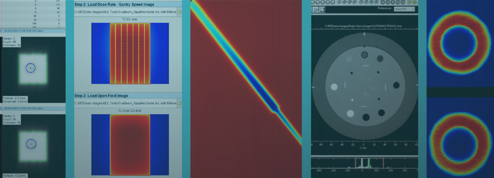
Fully-Automated Star Shot Analysis
RIT’s film Star Shot beam detection routine has been completely re-designed and now features a fully-automated interface with more robust and accurate detection algorithms. A few key highlights include:
- Full Automation: Increase your efficiency and accuracy with RIT's one-click analysis of Star Shot images. The new algorithm removes pre-processing from your workflow, allowing for quicker acquisition of results. Polarity, ROI, number of spokes, and spoke center are all automatically extracted from the image, then applied in the analysis.
- Visualized Spoke Detection: Newly-added beam center overlays on the beam profile plots provide a clear indication of the automated routine’s performance.
- Artificial Intelligence: The re-designed routine utilizes sophisticated AI algorithms for even more accurate and reliable measurements. The AI obtains a 99% accuracy rate of spoke detection in a wide range of Star Shot images.
- Manual Override Option: If desired, the routine will also allow you to easily modify the settings to fine-tune the automated analysis by hand.


Enhanced 3D Winston-Lutz (Isocenter Optimization)
- Newly-Added Clinical Angles: Automatically process a range of 3 to 16 EPID Winston-Lutz images for fast, sub-pixel level accuracy of isocenter position. This newly-enhanced test will allow physicists to perform daily or patient-specific analyses using specific clinical treatment angles, with a ball setup error reported for each test.
- Detailed PDF Report Customization: You may now customize your exported PDF reports, choosing to include or exclude plot and table data such as axis projection details, angle configurations, and optimization results, among others.
- Enhanced Axis Projection Table Display: For added convenience, the axis projection detail window now displays the data table, allowing you to quickly and easily reference your measurements.
MLC QA
CyberKnife® TG-135 QA: Fully-Automated M6 MLC Test
RIT's fully-automated “Garden Fence” MLC test, included in RIT Complete and RITG135, greatly increases the efficiency of MLC tests for CyberKnife units with a M6 multi-leaf collimator*. The MLC tests are consolidated into a single software package that will automatically crop the image, automatically align the image, perform automatic orientation of any images that are rotated or flipped, and automatically detect any leaves, eliminating any need for template files.
*According to experts at Accuray, you can save an average of 30 minutes per film.
CyberKnife® is a registered trademark of Accuray, Inc.


Elekta Leaf Speed Analysis
Efficiently perform automated, precision MLC QA for Elekta machines, with minimal setup. The new Leaf Speed Analysis routine measures the consistency and accuracy of the MLC leaf speeds as they traverse the imager. Similar to treatment deliveries, all measurements are made on an EPID, without the use of log files.
Patient QA
Automated Image Fill for Anthropomorphic Phantom QA
Use this newly-added function in the ‘Edit’ menu to automatically correct and fill any holes or cutouts in the image file. Perform patient QA with an anthropomorphic phantom for both calibrated and uncalibrated images. This feature supports a wide variety of anthropomorphic phantoms, including those used for CyberKnife® patient QA.
CyberKnife® is a registered trademark of Accuray, Inc.


Enhanced TomoTherapy® Registration
Easily perform exact dose comparisons for TomoTherapy patient QA. The newly-enhanced analysis uses a TomoTherapy plan, a dose map, and a film to determine position and dose accuracy using the red or green lasers. Coronal and sagittal slices may be analyzed using the following treatment planning systems from Accuray: TomoTherapy 5.1.3, Precision® 1.1.0, Precision® 2.0.0, and Precision® 3.0.0. Please note: This list has since been updated for the current version. For the most up-to-date list, please click here.
TomoTherapy® and Precision® are registered trademarks of Accuray, Inc.
Additional Accuray TPS Image Import Types
RIT software directly supports added Accuray treatment planning systems, including Accuray MultiPlan® and Precision® 3D DICOM file imports, used by clinical medical physicists to build optimized and precise plans for delivery on all Accuray systems.
MultiPlan® and Precision® are registered trademarks of Accuray, Inc.
Added RaySearch TPS Image Import Types
The RIT Family of Products software packages now directly support RaySearch 3D DICOM image file imports from the RayStation® treatment planning system - Version 8, used to build dynamic and adaptive delivery plans for a variety of treatments.
RayStation® is a registered trademark of RaySearch Laboratories AB.
Added PDF Report Export Options
Easily export the results of all patient QA measurements, including those performed in RIT's automated batch analysis feature, RunQueueA, to a customizable PDF report to display your most significant data.
Imaging QA/QC
Expanded Tolerance Customization
Utilize RIT’s newly-enhanced Tolerance Manager for increased customization capability. Use the Tolerance Manager to set completely custom values and pass/fail criteria for every measurement used in all automated phantom analyses. This tool provides the flexibility necessary to customize tolerances on a wide array of LINACs and imaging systems.

Gamma Camera Uniformity Analysis
Easily measure the Intrinsic Flood Field Uniformity (NEMA NU 1-2012) parameters in RIT software with a single click. Perform automated analysis of Integral Uniformity, Differential Uniformity, and Defective Pixels for modern, rectangular gamma cameras.

Universal CT Water Phantom Uniformity Analysis
Perform fully-automated, one-click analysis of any CT water uniformity phantom. This test allows you to easily adjust the size and position of the analysis areas to match any uniformity phantom. This routine is perfect for daily axial and helical uniformity measurements.
Other
Flatbed Non-Uniformity Correction
Now available to customers in the U.S., RIT’s Flatbed Non-Uniformity Correction feature corrects for flatness and uniformity variations in scanners, corrects for non-uniformities in EBT film to improve the film's dosimetric accuracy, and provides you with the option to automatically-generate a calibration file. This feature is now available in the following software packages: RIT Complete, RIT Classic, RITG142, and RIT Film.


New Licensing Options: Local License Server
In addition to floating and node-locked licenses, RIT now offers 'Local License Server' as an option for your software license management. With no internet required, the local license server is sent through your facility’s network and is tied to a specific server. This new option will allow users to have floating license capability, local to their facility’s network.