

An impressive range of products
Radiological Imaging Technology
Machine QA MLC QA Patient QA Imaging QA
What's New in V6.6 & Earlier
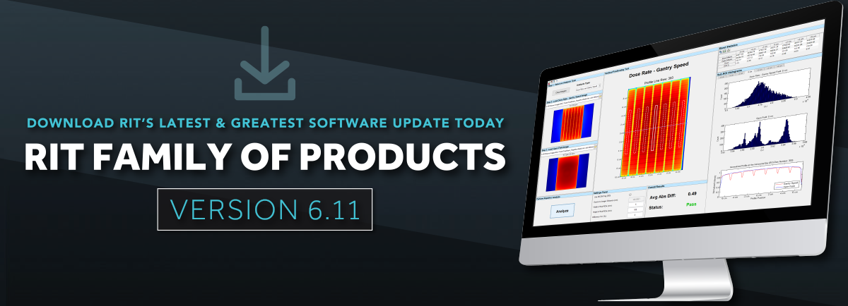
THE RIT FAMILY OF PRODUCTS: Version 6.6 & Earlier
Version 6.6 of the RIT Family of Products (Radia Version 1.10) was released on June 27, 2017.
Machine QA
3D Winston-Lutz Isocenter QA
The RIT Family of Products come equipped with an automated 3D Stereotactic Alignment routine to give you a fast and accurate measurement of isocenter position. Using only a set of EPID Winston-Lutz images, the RIT system will calculate isocenter displacement, determine wobble around the isocenter, and suggest couch position adjustments to optimize your system’s accuracy. Eliminate your need for films, and increase your accuracy by using the all new Virtual Starshot, reconstructed using a set of Winston-Lutz images!*
Updates from Version 6.6:
- Updated 3D Winston-Lutz analysis interface makes relevant measurements more visible and intuitive.
- Analysis automatically determines ball setup error, providing analysis results both with and without setup error contribution.
- Use Virtual Star Shot* with ANY combination of angles in its dedicated window.
- The couch optimization routine includes tabs for intuitive visualization of adjustment outcome.
- Organize measurement table results to pinpoint minimum and maximum values.
- PDF report includes footnote definitions to easily interpret analysis results.
Updates from Version 6.5:
- All collimator angles are supported.
- “Old Varian” and Siemens coordinate systems are supported.
- Optimization of region-of-interest routine for automatic field size detection.
- Improvements to the Axis Projection screen
Updates from Version 6.4:
- Eliminate your need for films, and increase your accuracy by using the Virtual Starshot, reconstructed using a set of Winston-Lutz images!*
Updates from Version 6.2:
- Added the ability to choose amongst a number of alternate coordinate systems within the routine, so that the results are relevant to your system’s configuration.
Updates from Version 6.1:
- RIT’s version of this classic test allows you to use 3-10 images and not only gives you an accurate measurement of isocenter displacement but also an error estimate to determine the wobble around the isocenter.
*US Patent 9192784, JP Patent 6009705, CA Patent 2918045, and other International Patents pending

RITG148+: Helical TomoTherapy®

Updates from Version 6.6:
- PDF reports for all analysis routines
- Vidar TIFF Export
- Cheese phantom enhancements:
- New geometric distortion measurement
- Improvements to noise and uniformity analysis - Static Gantry Angle analysis enhancements
- Crop images with Pin-Prick and Erase tool
Updates from Version 6.4:
Added tests include:
- Interrupted Treatment
- Image Quality Tests (Cheese Phantom) which include: Noise, uniformity, resolution, and contrast
- Automated QA testing
- Y-jaw divergence/beam centering
- Y-jaw/gantry rotation plane alignment
- Treatment field centering
- MLC alignment test
- Couch translation/gantry rotation
- Laser localization
- Image quality tests (Cheese Phantom)
- Built-in trending and reporting with RITtrend™
- Helical and Static Gantry Angle Consistency tests
RITG135: Full CyberKnife® and All Robotic Radiosurgery QA
(Version 6.6)
Developed in accordance with TG-135 specifications
- End-to-End Test
- AQA Test
- IRIS Test
- Laser Coincidence Test
- Fully Automated MLC Test for the M6 Collimator
Beam Measurements
- Enhanced Radiation/Light Field Coincidence test (Version 6.6)
- New Asymmetrical Light/Radiation field test (Version 6.5)


Added TG-142 Tests Using Your EPID (V6.3)
![]()
One Comprehensive Software Solution:
Patient QA
2D Array Analysis File Types (Version 6.6)
- 2D Array analysis now supports 3D DICOM and .mat image files.
Improved Radiochromic Non-Uniformity Correction (Version 6.6)
- This routine reduces effect of film uniformity concerns to improve QA results.
Welcome to the Family: RIT Film (Version 6.5)
- Full suite of film dosimetry routines including patient and machine QA
IMRT (Patient QA) Analysis (Version 6.5)
- Enhancements to 2D Array IMRT Analysis routines

MLC QA
Elekta Hancock Test (V6.0)
Updates from Version 6.6:
- The enhanced Hancock Test offers the ability to utilize just two images for analysis, significantly reducing the time needed to perform this test.
- No more going into the treatment room to manually extend the image panel; all images can be taken without leaving the operating console.
Updates from Version 6.3:
- This version introduced a "leaves only" version of the popular Hancock test with even greater accuracy and simplicity. Using 4 simple images the test accurately measures absolute leaf position with respect to machine isocenter.
- This is for both the newer 160 leaf Agility MLC and the older.
- The 80 leaf MLCi2 head are supported for this test.
Updates from Version 6.2:
- Added independent control over jaw and leaf thresholding to give users more control over the analysis, along with a number of algorithm choices to match your precise situation.
Updates from Version 6.1:
- This unique test that measures both leaf position and jaw setback on Elekta machines has had a number of new pattern options added to make it even more flexible and robust.


Picket Fence Test (Version 6.4)
- Support for Varian 40x40 EPID for both Millennium 120 and Millennium 120 HD MLC
RIT EPID MLC And RapidArc Tests 0.2, 1.1, 1.2 (V6.2)
- Added HD MLC versions of all these tests so that you can enjoy the same speed and accuracy on analyzing your HD picket fence patterns as you do with your Millennium MLCs.
- Automated the recognition of the pixel value polarity, so there is no more requirement to scale images from your True Beam machines.
Auto MLC Routine for EPID (V6.0)
- Automatically compensates for SID and imager offset to find FOV intelligently – no typing in SID or taking ROIs
- New leaf position and FWHM distribution plots
Imaging QA/QC
Phantom Analysis
Updates from Version 6.6:
- Added 6-Degree-of-Freedom Couch Test, utilized in parallel with the ISOCube™ to verify the mechanical accuracy of couch motion
- Added support for analysis of the Catphan® 604 phantom and Penta-Guide phantom
- Ability to perform QC on Gamma Knife Icon’s integrated CBCT unit using the Catphan®
Updates from Version 6.5:
- Catphan® 404 materials specifications have been updated to allow for much easier customization of HU constancy targets. Nominal slice thickness calculation now includes frame averaging. A water vial can now be used in the top “air” plug for more complete analysis.
- Improvements have been made to the TOR-18FG Module, which include defaults in settings now offering compatibility to more images without adjustments, and improvements to the graphs.
- The XVI Sagittal Scaling Module now allows for adjustments of vertical and axial offsets to support non-standard scanning parameters. Enhancements have also been made to the slice averaging routines provide updated DICOM headers after averaging.
Updates from Version 6.2:
- Added the capability to designate which collimator was used in the image acquisition and adjust the pass/fail criteria appropriately to reflect differences in expected image quality between the s10 collimator (typically used in commissioning) and the wider s20 collimator typically used in periodic QA.
- Added the Sagittal Geometry test from the XVI acceptance test suite. Simply open an image from the folder with the complete CT image set and will create a sagittal reconstruction and measure the sagittal geometric accuracy in accordance to the XVI acceptance specifications.
Updates from Version 6.0:
Imaging QA capabilities were added to RIT software for automated phantom analysis.
- Meet all of your TG142 Imaging QA requirements
- Supports most common phantoms for CT, CBCT, kV and MV imaging on all LINAC manufacturers’ equipment
- Fast, totally automated analysis of individual images or image sets
- Automated retrieval of multiple images


Cerberus File Watcher (V6.1)
- Have you ever wished that your RIT system could monitor a folder and process incoming files without you lifting a finger or even being present? You can now with RIT’s Cerberus system.
- Configure the system to watch for the specific files you want to process and set it loose.
- Perfect for situations where images are being generated in multiple locations.
- Send them to a watched folder and have them analyzed automatically.
Other Enhancements
- Use RunQueueC’s improved capability to handle more files for analysis. (V6.6)
- Utilize several new enhancements to Cerberus functionality, including the DICOM tag filter, which now provides more robust search functionality. (V6.6)
- Added an ROI tool to the Imaging QA interface. (V6.2)
- Added orthogonal rotation ability to the Image QA interface. (V6.2)
- Easier access to the analysis preferences. (V6.2)
Other Features
Global Preferences Manager (V6.6)
- Create custom preferences profiles for each user, machine, or facility location.
- Easily manage all preferences and settings in one, centralized location.

PDF Reports for Every Analysis Routine (V6.6)
- Easily export ANY analysis routine as a PDF report with a single click!
- Customize PDF reports to display specific information you find most important.
Enhanced Editing (V6.6)
- Imported images can be edited utilizing Non-Cropping Rotation and Resize tools.
- Non-Cropping Rotation and Resize tools support an extensively-increased range of file types.
- Pin-Prick and Erase tools now have the ability to crop images.
64-Bit Application (V6.5)
- If you run the 64-bit version on a computer with 16Gbytes of RAM, it is blazing fast! Why wait?
- Take confidence knowing that this updated version provides exactly the same results and is held to the same standard of quality RIT is known for.
Directory Browser and DICOM Directory Browser for the RIT Family of Products (V6.4)
Easily organize and view test image thumbnails with the new Directory Browser and DICOM Directory Browser. Images can be sorted with quick and easy viewing of DICOM header information. Double-click any image to transfer to the main RIT interface for analysis. Ideal for use with the RIT TG-142 Patient.
RITtrend™ Statistical Database Analysis
Updates in Version 6.4:
- The RITtrend™ database has been converted to an SQL Server database. This seamless transition will allow for greater long term flexibility, speed, stability and robustness going forward.
- Be notified when added data exceeds customizable warning and failure limits or SPC control limits via email alerts.
- All your common tasks such as querying, managing tolerances and SPC limits have been greatly simplified reducing clicks and time by more than 80%. The new interface allows you to efficiently visualize and manage large homogenous datasets generated from many tests in a simple and intuitive manner.
- Locate your database anywhere on your local machine, network, or the cloud.
- Build customized reports for compliance with TG142 requirements.
- Improved options for finding the data you need.
Updates in Version 6.2:
- Improved speed with optimized load times on large databases
- Querying over designated date ranges easier and simpler.
Updates in Version 6.0:
The RITtrend™ tracking and trending feature was added to the RIT Family of Products.
- Establish and manage baselines and specifications
- Compare measurements across databases
- Design and store custom queries for any measurement
- TG-142 Report - one custom report to track all your requirements
