RIT Complete

Medical Physics' most advanced, comprehensive radiation therapy quality assurance software package, combining Machine QA, MLC QA, Patient QA, and Imaging QA all in one.
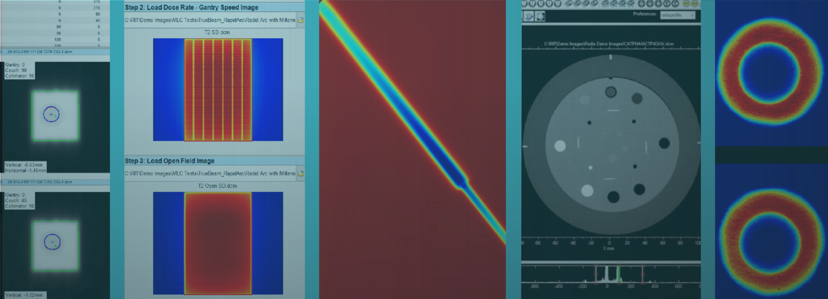
Streamline your quality assurance measurements with the all-in-one automated software solution, RIT Complete. RIT Complete consolidates all of RIT’s innovative therapy products into one comprehensive QA solution, with all the machine, MLC, and patient QA included in the RIT Classic product, with the addition of Imaging QA for a variety of therapeutic phantoms. RIT Complete utilizes a combination of powerful, robust routines in a user-friendly interface to maximize the efficiency and precision of your measurements. For added convenience, you can easily export your analysis data as customizable reports or to the RITtrend™ statistical database for large-scale tracking and trending over time.
Machine QA
RIT Complete provides extensive machine QA capabilities with a full suite of measurements in accordance with TG-142 (including MPPG 8.b), TG-148, and/or TG-135 recommendations. RIT’s automated routines allow you to perform daily, monthly, and annual QA with efficiency and accuracy, all while giving you confidence knowing your delivery performance is optimized.
Daily, Monthly, and Annual Machine QA for LINACs
- Enhanced 3D Winston-Lutz (Isocenter Optimization) with Virtual Star Shot*
Automatically process a set of EPID Winston-Lutz images for a fast and accurate measurement of isocenter position. RIT’s version of this test allows you to use 3 to 16 images, and provides error estimates for ball setup and wobble around isocenter. Increased angle flexibility allows you to mimic more clinically relevant angles to determine isocenter at specific treatment configurations. *US Patent 9192784, JP Patent 6009705, CA Patent 2918045, and other international patents pending. - Stereotactic Alignment (2D Winston-Lutz) Test
- Stereotactic Cone Profiles
- Field Alignment
- Gibbs Cone Analysis

TomoTherapy® Measurements
- Beam Planarity and Jaw Twist
- Overhead Laser Position Tool
- Import TomoTherapy® DICOM Film Files
- Import TomoTherapy® Calibration Files
- Static Gantry Angle Tool
- Helical Gantry Angle Tool
- Field Center vs. Jaw Setting Tool
- Couch Translation/Gantry Rotation
- Interrupted Treatment
- MLC Center of Rotation Tool
- IGRT Alignment
- Vidar TIFF Export
TomoTherapy® is a registered trademark of Accuray, Inc.

Beam Measurements
- Fully-Automated Star Shot Analysis
RIT’s enhanced Star Shot beam detection routine has a fully-automated interface with robust and highly accurate artificial intelligence algorithms. Polarity, ROI, number of spokes, and spoke center are automatically extracted from the image and applied in - Radiation/Light Field Coincidence
- Asymmetric Field/Matchline
- Electron Energy (TG-25)
- Quick Flatness and Symmetry
- Water Tank Beam Measurement Analysis
- Depth Dose, Cross, and Orthogonal Profiles
- Isodose Contours
- Image Histogram
- 3D Dose Profile

CyberKnife® & All Robotic Radiosurgery Measurements
- End-to-End Test
- AQA Test
- Laser Coincidence Test
- Iris Test
- M6 MLC Test
CyberKnife® is a registered trademark of Accuray, Inc.

MLC QA
As part of RIT’s comprehensive Machine QA capabilities, RIT Complete maximizes the precision of quality assurance for multi-leaf collimators on any linear accelerator, including Varian LINACs, Elekta LINACs, CyberKnife® M6 MLC, and more. The software allows users to track MLC performance over time and have confidence that patient treatments are proceeding as planned.
Varian LINAC
- EPID Picket Fence Test
This routine automates the classic picket fence test. - Automated Varian RapidArc® Tests
Images may be taken at any distance from EPID, Film, or CR Images. This includes: Tests 0.1, 0.2, 1.1, 1.2, 2 and 3. RIT’s RapidArc QA routines support the Millennium 120, HD120 MLC, and Halcyon® MLC models. - Varian Leaf Speed Test
Without the use of log files, this test measures the consistency and accuracy of Varian MLC leaf speeds as they move across an imager. - Varian Halcyon® MLC Analysis
Perform a picket fence or comprehensive RapidArc® analysis of the Halcyon® MLC.

Elekta LINAC
- Hancock Tests for Elekta Machines
The Hancock Tests (2-Image Test, 4-Image Test, and With Backup Jaw Test) use the Elekta iView™ imager to automatically measure leaf position vs. isocenter position, and jaw leaf setback measurements. - EPID Picket Fence Test
This routine automates the classic picket fence test. - Elekta Leaf Speed Test
This test aligns two images to analyze the consistency of the leaf speed for both Elekta iView™ and Agility™.

CyberKnife® MLC
- CyberKnife M6 MLC QA
Perform a fully automated "Garden Fence" MLC test for the M6 multi-leaf collimator. The software will (1) automatically crop the image; (2) automatically align the image; (3) perform automatic orientation of any images that are rotated or flipped; (4) automatically detect any leaves, eliminating the need for template files; and (5) perform the analysis. According to experts at Accuray, you can save an average of 30 minutes per film with RIT software.

Other MLC Tests
- Bayouth MLC Analysis
Analyze MLCs that require leaf gaps between banks. - TomoTherapy® MLC Leakage Analysis
Fully automate the MLC leakage calculation specified by Accuray, which can significantly increase the uniformity of your measurements. - Additional MLC Tests
These include: TG-50 Picket Fence Test, MSK Leaf Test, Varian DMLC Test Patterns, and MLC Transmission analysis.
iView™ and Agility™ are trademarks of Elekta AB. RapidArc® and Halcyon® are registered trademarks of Varian Medical Systems, Inc.
Patient QA
RIT was the first company to develop software for intensity-modulated radiation therapy (IMRT) analysis, and RIT continues to lead the industry with automated patient QA routines in RIT Complete. The software provides clinical physicists with the tools needed to analyze this advanced cancer treatment, ensuring rapid results without compromising measurement precision.
TomoTherapy® Registration
Easily perform exact dose comparisons for TomoTherapy patient QA. This innovative wizard uses a TomoTherapy plan, a dose map, and a film to determine position and dose accuracy, using the red or green lasers.This automated technique extends standard registration methods, as the isocenter location is not required to be in the center of the exposed dose region. Coronal or Sagittal slices may be analyzed, and the routine allows a structure to display as an overlay ROI. Conveniently select a specific structure (e.g., PTV) from any of the plan structures and display the Structure Outline on the image, allowing your analysis to be limited to the structure of your choosing, plus a user-defined margin. Then, use RIT’s Patient QA routines to measure your results (Gamma, Subtraction, DTA, or one of 28 other analysis routines) within the ROI. Utilize RIT’s patented Plan-Based Calibration technology with this routine. TomoTherapy® and Precision® are registered trademarks of Accuray, Inc.

IMRT, IGRT & RapidArc®/VMAT Analysis
- Gamma Analysis
- Distance-to-Agreement (DTA)
- Profiles
- Van Dyk’s Analysis
- Subtraction
- Composite Analysis
- Isodose Curves
- Addition
- Centroid Measurement
- IGRT Alignment
- IMRT Fine-Tune Registration
- Bilinear Interpolation & Non-Cropping Rotation
- Proportion Passing Plot
- Save Case Files from IMRT Analysis Toolbars
- Save & Restore IMRT Layouts
- Plan-Based Calibration
(Patents: EP 1683546, CA 2567197, JP 4366362, JP 4838161, US 7024026, US 7233688, US 7639851, and US 7680310.)

2D Detector Array Analysis
- Import from 2D Arrays: Ion Chamber Arrays from IBA, PTW, and MapCHECK® diode arrays are supported. IMRT analysis routines have been revised to handle sparse data. Results can be saved as a RIT Array Case file.
Automated Image Fill for Anthropomorphic Phantom QA
- Use this function to automatically correct and fill any holes or cutouts in the image file. Perform patient QA with an anthropomorphic phantom for both calibrated and uncalibrated images.
Patient QA Image Registration
-
Simultaneously perform fully-automated registration control point positioning in both traditional and RunQueueA (automated batch analysis) IMRT. Template-based registration may also be performed.
Dose Calibrations
- Perpendicular dose calibration
- Parallel dose calibration
- MLC calibration technique
(Patents: EP 1318857, CA 2418232, JP 3817176, US 6675116, US 6934653, and US 7013228) - iView™ calibration
- Kodak CR (perpendicular and spatial calibrations),
- Optical density (OD) calibration
- Daily output factor adjustment for calibration curves.
- Scanner Spatial Calibration
The spatial calibration is not a dose conversion, but rather a means to determine the exact pixel size for the Vidar film scanner or flatbed scanner. This gives you the most accurate distance measurements. - PDD Table Editor
- Calibration File Merge
MapCHECK® is a registered trademark of Sun Nuclear Corp.
Imaging QA
RIT Complete offers extensive Imaging QA/QC capabilities, from specific routines following task group recommendations, to a full suite of one-click, instant phantom analyses of therapeutic and diagnostic images. RIT Complete comes equipped with tracking, trending, tolerance customization, and hands-free automation tools designed to streamline your workflow.
Custom Tolerance Management
Use the Tolerance Manager to set tolerance values and pass/fail criteria for every measurement used in all automated phantom analyses. This tool provides the flexibility necessary to customize tolerances on a wide array of LINACs and imaging systems:
- Conveniently set and toggle between tolerances in an easy-to-use panel, located in the main interface, and displayed in every phantom analysis routine.
- Set your own PASS/FAIL criteria for analyses and their corresponding reports, allowing for complete customization in all imaging QA analyses.
- Preference profiles and tolerances are set on a per-machine basis. They can be precisely-tailored to each individual machine in use.
- Transfer all PASS/FAIL tolerance data to the RITtrend™ database for statistical tracking and trending.

CATPHAN®, kV and Cone Beam Module
- Varian 504 CATPHAN
- Varian 604 CATPHAN
- Elekta 503 CATPHAN
- Siemens Image Quality Phantom
Electron Density Module
- Cheese Phantom
- CIRS 062, 062A, 062M (Cone Beam)
- Gammex 467
kV/MV Module
- Standard Imaging ISOCube
- Penta-Guide

EPID QC Module
- RIT EPID
(EPID QA Phantom - manufactured and sold by Standard Imaging.) - PTW EPID
- Las Vegas EPID
- Standard Imaging QC-3
DR/CR kV Module
- IBA Primus® L
- PTW NORMI® 4
(20x20cm and 30x30cm) - Leeds TOR-18 FG
- Standard Imaging QC-kV1
Base Module
- DICOM Image Viewer
- DICOM Tag Viewer
- Image Import: DICOM Directory Browser, Open Directory Browser, DICOM File Filter
- Manual measurements: distance, angle, profile, histograms, round and square ROI, and ASCII output of profiles
- Output formats: Print, PDF export, Excel export with custom templates
- Monitor Image Quality
- Image transfer
Other Measurements
Film Dosimetry for QA
- Radiochromic and Radiochromic Film with Vidar and Flatbed Scanners
- Flatbed Non-Uniformity Correction
- Radiochromic Film Uniformity Correction
- Automated 21-Point Film Processor Correction
(Patents: EP 1252550, CA 2396952, JP 3817176, and US 6528803) - Sensitometry
(Patents: EP 1252550, CA 2396952, JP 3817176, and US 6528803) - 2D Scanner Spatial Calibration for Vidar and Flatbed Scanners
- Generic Image File Import
- Vidar Advantage Pro 180° Correction
- Vidar Scanner Interface (Vidar Scanner Control Center)
Computed Radiography (CR)
- 3D CR Flatness and Uniformity Correction
- Kodak CR Perpendicular Calibration
- Kodak CR Spatial Calibration
EPID QA
- Import EPID Images for QA
- EPID Calibration
- Elekta iView™ Calibration
- Scale DICOM Images from Varian EPIDs
General Features
- Image Compositor
- DICOM Anonymizer
- Pin Prick, Erase, and ROI Tools
- PDF reports for every analysis routine
- Cloud-based software licensing
- Support of 3D gels and solids
Built-In Automation Features in RIT Complete

RIT Complete includes RITtrend™ – the all-in-one statistical database tracking solution, that stores data for all your department's measurements. Set your own specifications and RITtrend automatically analyzes process control limits on equipment analyzed with RIT software. The multi-source data manager gives you even more control for reporting on your entire medical physics program. RITtrend redefines database recording into a major tool for analysis and record-keeping in your department.
RITtrend™ is a trademark of Radiological Imaging Technology, Inc.

RIT Complete includes RunQueueA – the automated patient QA batch analysis feature. Automate your patient QA by setting up scripts for your repetitive patient QA/IMRT workflows. Perform automated matching and sorting of reference and target images simultaneously. Easily export your results to a customizable PDF report to display your most significant data.

RIT Complete includes RunQueueC – the fully automated batch phantom analysis tool. RunQueueC is an automated feature included in all RIT software packages with imaging QC capabilities. With one click, easily perform analysis on any number of images. RunQueueC will automatically generate analysis reports in a designated folder and export data to the RITtrend database for statistical tracking and trending over time.

RIT Complete includes Cerberus – the hands-free batch phantom analysis tool. Cerberus constantly operates in the background of your workstation, automatically monitoring folders and pin-pointing specific files to process and analyze. It can match many set criteria, such as file naming patterns, DICOM tag matches (up to 4 DICOM tags may be used), or file types. Cerberus automatically performs analyses, generates reports, and shares data to RITtrend, using specific parameters set in your customized preference profiles (including tolerances) to analyze the images for all of your machines.
Upgrade YOUR PRODUCT today!
RIT also offers the RIT Complete Diagnostic package. The RIT Complete Diagnostic product package allows for automated ACR CT and ACR Large MRI phantom analysis, in addition to all the Patient, Machine, MLC, and Imaging QA included in the standard RIT Complete package. Simple, easy-to-use tools provide complete scoring of all phantom parameters, with RIT's characteristic precision and accuracy. For more information, please contact RIT Sales.
719-590-1077, Opt. 4
 All of RIT’s therapy products in one convenient and comprehensive package |
||||
 The original RIT product combining patient and machine QA |


