
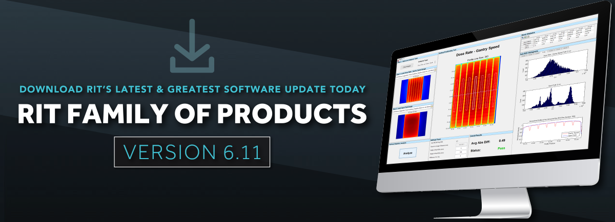

An impressive range of products
Radiological Imaging Technology
Machine QA MLC QA Patient QA Imaging QA
RIT Film
![]()
RIT's software product for film dosimetry with Patient QA and basic Machine & MLC QA
The RIT Film software package offers a full suite of film dosimetry routines, including Patient QA and basic Machine QA. Easily streamline your Patient QA with RunQueueA, RIT’s automated batch analysis feature, and easily export any analysis routine as a PDF report with a single click.
Patient QA
RIT Film’s Patient QA (IMRT) routines are designed for the comparison of a patient’s phantom plan from the user’s treatment planning system and the QA image captured by film exposed to that phantom plan. The software conveniently allows for comparing a plan and film, a plan to plan, and/or a film to film.
IMRT & VMAT Analysis
- RIT's Patented Plan-Based Calibration
Make quick, relative comparisons between any dose map and your EPID, film, and CR images.
(Patents: EP 1683546, CA 2567197, JP 4366362, JP 4838161, US 7024026, US 7233688, US 7639851, and US 7680310.)
- Gamma Function
- Distance to Agreement (DTA)
- Profiles
- Van Dyk's Analysis
- Subtraction
- Composite Analysis
- Isodose Curves
- Addition
- Centroid Measurement
- IMRT Automatic Fine-Tune Registration
- Automated Registration
- Proportion Passing Plot
- Patient QA Image Registration
Simultaneously perform fully-automated registration control point positioning in both traditional and RunQueueA IMRT. Template-based registration may also be performed.

2D Detector Array Analysis
- Import from 2D Arrays: Ion Chamber Arrays from IBA, PTW, and MapCHECK® diode arrays are supported. IMRT analysis routines have been revised to handle sparse data. Results can be saved as a RIT Array Case file.
MapCHECK® is a registered trademark of Sun Nuclear Corp.
Automated Image Fill for Anthropomorphic Phantom QA
- Use this function to automatically correct and fill any holes or cutouts in the image file. Perform patient QA with an anthropomorphic phantom for both calibrated and uncalibrated images.
Calibration
- RIT's Patented MLC Calibration Technique
(Patents: EP 1318857, CA 2418232, JP 3817176, US 6675116, US 6934653, and US 7013228.) - Perpendicular Dose Calibration
- Parallel Dose Calibration
- Optical Density (OD) Calibration
- 2D Scanner Spatial Calibration for both Vidar and Flatbed Scanners
- PDD Table Editor
- Calibration File Merge
Partial Machine & MLC QA
Machine QA & Beam Measurements
- Fully-Automated Star Shot Analysis
RIT’s enhanced Star Shot beam detection routine has a fully-automated interface with robust and highly accurate artificial intelligence algorithms. Polarity, ROI, number of spokes, and spoke center are automatically extracted from the image and applied in - Radiation/Light Field Coincidence
Radiation/Light Field EPID images may be analyzed without applying a calibration file. You can also have custom field sizes (not just 5cm, 10cm, 15cm, and 20cm), and use a BB or pinprick for center location, or use an L-Rad phantom. - Depth Dose, Cross, and Orthogonal Profiles
- Electron Energy (TG-25)
- Quick Flatness and Symmetry
- Isodose Contours
- Stereotactic Alignment (2D Winston-Lutz) Test
- Stereotactic Cone Profiles
- Field Alignment
- Import TomoTherapy® Calibration Files
MLC QA
- Bayouth MLC Analysis
Analyze MLCs that require leaf gaps between banks. - Generic Picket Fence Test (Memorial Sloan Kettering Picket Fence Pattern)
- MLC Transmission Analysis (TG50 Recommended)
- TG50 Picket Fence
- Varian DMLC Test Pattern Analysis
- Test 1: Picket Fence
- Test 2: Synchronized Segmented Stripes
- Test 3: Non-Synchronized Segmented Stripes
- Test 4: X Wedge Single Image & Composite
- Test 5: Y Wedge Single Image & Composite
- Test 6: Pyramid Single Image & Composite
- Test 7: Complex A
- Test 8: Complex B
- Test 9: Continuous Stripes
Other Measurements
Film Dosimetry for QA
- EBT3 and Flatbed Scanner Correction
This advanced feature corrects for flatness and uniformity variations in scanners, corrects for non-uniformities in EBT3 film to improve the film’s dosimetric accuracy, and provides you with the option to automatically-generate a calibration file. - Radiochromic and Radiochromic Film with Vidar and Flatbed Scanners
- Flatbed Non-Uniformity Correction
- Radiochromic Film Uniformity Correction
- Automated 21-Point Film Processor Correction
(Patents: EP 1252550, CA 2396952, JP 3817176, and US 6528803) - Sensitometry
(Patents: EP 1252550, CA 2396952, JP 3817176, and US 6528803) - 2D Scanner Spatial Calibration for Vidar and Flatbed Scanners
- Generic Image File Import
Import generic JPEG, TIFF, and bitmap image files from sources other than a VIDAR Scanner, giving you increased flexibility in your workflow. - Vidar Advantage Pro 180° Correction
- Vidar Scanner Interface (Vidar Scanner Control Center)

General Features
- DICOM Anonymizer
- Pin Prick, Erase, and ROI Tools
- PDF reports for every analysis routine
- Cloud-based software licensing
- Support of 3D gels and solids
Built-In Features in RIT Film

As a built-in feature, RIT Film includes RunQueueA – the automated patient QA batch analysis feature. Automate your patient QA by setting up scripts for your repetitive patient QA/IMRT workflows. Perform automated matching and sorting of reference and target images simultaneously. Easily export your results to a customizable PDF report to display your most significant data.